CT Scanning
CT SCANS
Computer Tomography (CT) was first developed in the early 70's as a method of imaging using Xrays.
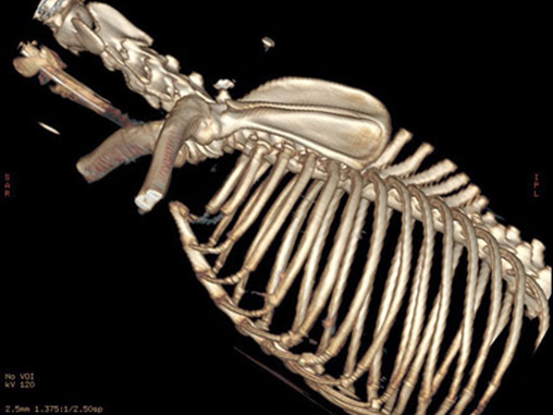
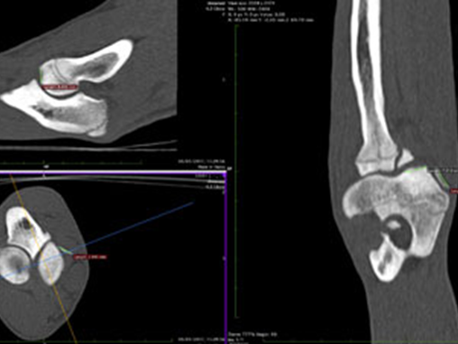
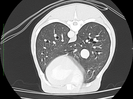
Because the technique uses a very fine beam of Xrays detected by a fine array of sensors a 3D picture can be built up of the subject being examined. There are three types of CT, helical, linear and cone beam, helical and linear provide the greatest image quality allowing detailed imaging of patients as large as a Great Dane’s chest to as small as a kitten’s foot.
The inherent contrast afforded by the differences between, air, bone, fat and soft tissue is also enhanced by the use of Iodinated intravenous or intra arterial contrast (intravenous being the most common) and so very fine detail of tomography, internal architecture and vascular supply and drainage is possible for bones, the abdomen, the chest, head and neck in fact almost every body part. Only brain, spinal cord and muscular tendinous injuries are areas where MRI may provide superior imaging.
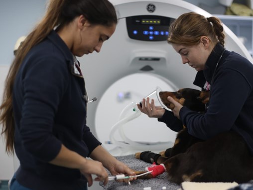
CT scanning is as quick as taking standard xrays and has proved to be invaluable to the improved diagnostics for orthopaedic disease and injuries for cats and dogs, especially in elbow dysplasia and patella luxation where the improved diagnostics are making our treatments more accurate with improved functional outcomes and reduced complications.
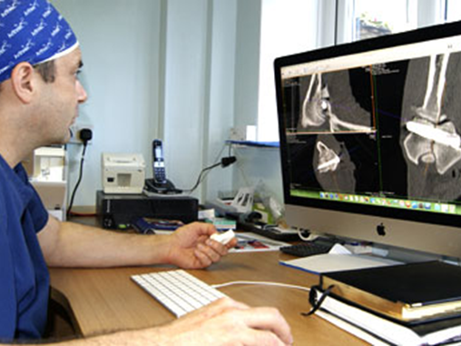
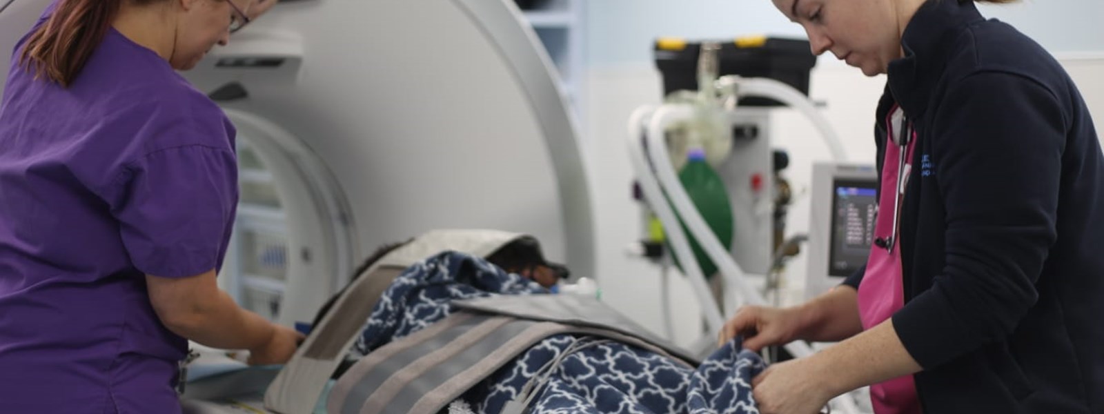
Having this facility on site also allows us to provide these benefits to your pet when appropriate.